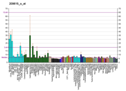
PAK1
PAK1 (ингл. ) — аксымы, шул ук исемдәге ген тарафыннан кодлана торган югары молекуляр органик матдә.[26][27]
Искәрмәләр
үзгәртү- ↑ 1,0 1,1 UniProt
- ↑ 2,00 2,01 2,02 2,03 2,04 2,05 2,06 2,07 2,08 2,09 2,10 2,11 2,12 2,13 2,14 2,15 2,16 2,17 2,18 2,19 2,20 2,21 2,22 2,23 2,24 2,25 2,26 2,27 2,28 2,29 2,30 2,31 2,32 2,33 2,34 2,35 2,36 2,37 2,38 2,39 2,40 2,41 2,42 2,43 2,44 2,45 2,46 2,47 2,48 2,49 2,50 2,51 2,52 2,53 2,54 2,55 2,56 2,57 2,58 2,59 2,60 2,61 2,62 2,63 2,64 2,65 2,66 2,67 2,68 GOA
- ↑ Knaus U. G. A p21-activated kinase-controlled metabolic switch up-regulates phagocyte NADPH oxidase // J. Biol. Chem. / L. M. Gierasch — Baltimore [etc.]: American Society for Biochemistry and Molecular Biology, 2002. — ISSN 0021-9258; 1083-351X; 1067-8816 — doi:10.1074/JBC.M206650200 — PMID:12189148
- ↑ 4,0 4,1 4,2 Koh C., Tan E., Manser E. et al. The p21-activated kinase PAK is negatively regulated by POPX1 and POPX2, a pair of serine/threonine phosphatases of the PP2C family // Curr. Biol. — United Kingdom: Cell Press, Elsevier BV, 2002. — ISSN 0960-9822; 1879-0445 — doi:10.1016/S0960-9822(02)00652-8 — PMID:11864573
- ↑ 5,0 5,1 5,2 G Wang, Q Zhang, Y Song et al. PAK1 regulates RUFY3-mediated gastric cancer cell migration and invasion // Cell Death Dis. — London: Nature Publishing Group, 2015. — ISSN 2041-4889 — doi:10.1038/CDDIS.2015.50 — PMID:25766321
- ↑ Ijuin T. Glucose-regulated protein 78 (GRP78) binds directly to PIP3 phosphatase SKIP and determines its localization // Genes Cells / M. Yanagida — Wiley-Blackwell, 2016. — ISSN 1356-9597; 1365-2443 — doi:10.1111/GTC.12353 — PMID:26940976
- ↑ 7,0 7,1 7,2 7,3 7,4 7,5 7,6 GOA
- ↑ 8,0 8,1 8,2 8,3 Brown J. L., L Stowers, M Baer et al. Human Ste20 homologue hPAK1 links GTPases to the JNK MAP kinase pathway // Curr. Biol. — United Kingdom: Cell Press, Elsevier BV, 1996. — ISSN 0960-9822; 1879-0445 — doi:10.1016/S0960-9822(02)00546-8 — PMID:8805275
- ↑ Wang J., Wu J., Wang Z. Structural insights into the autoactivation mechanism of p21-activated protein kinase, Structural Insights into the Autoactivation Mechanism of p21-Activated Protein Kinase // Structure / C. D. Lima — Cell Press, Elsevier BV, 2011. — ISSN 0969-2126; 1878-4186 — doi:10.1016/J.STR.2011.10.013 — PMID:22153498
- ↑ Rayala S. K., Bert W O'Malley Signaling-dependent and coordinated regulation of transcription, splicing, and translation resides in a single coregulator, PCBP1 // Proc. Natl. Acad. Sci. U.S.A. / M. R. Berenbaum — [Washington, etc.], USA: National Academy of Sciences [etc.], 2007. — ISSN 0027-8424; 1091-6490 — doi:10.1073/PNAS.0701065104 — PMID:17389360
- ↑ Knaus U. G. Human p21-activated kinase (Pak1) regulates actin organization in mammalian cells // Curr. Biol. — United Kingdom: Cell Press, Elsevier BV, 1997. — ISSN 0960-9822; 1879-0445 — doi:10.1016/S0960-9822(97)70091-5 — PMID:9395435
- ↑ 12,0 12,1 12,2 12,3 Zegers M. M., Chernoff J. Pak1 and PIX regulate contact inhibition during epithelial wound healing // EMBO J. — NPG, 2003. — ISSN 0261-4189; 1460-2075 — doi:10.1093/EMBOJ/CDG398 — PMID:12912914
- ↑ G Wang, Q Zhang, Y Song et al. PAK1 regulates RUFY3-mediated gastric cancer cell migration and invasion // Cell Death Dis. — London: Nature Publishing Group, 2015. — ISSN 2041-4889 — doi:10.1038/CDDIS.2015.50 — PMID:25766321
- ↑ Talukder A. H., Q Meng, R Kumar CRIPak, a novel endogenous Pak1 inhibitor // Oncogene — NPG, 2006. — ISSN 0950-9232; 1476-5594 — doi:10.1038/SJ.ONC.1209172 — PMID:16278681
- ↑ Curtis I. D. Analysis of the subcellular distribution of avian p95-APP2, an ARF-GAP orthologous to mammalian paxillin kinase linker // Int. J. Biochem. Cell Biol. — Elsevier BV, 2002. — ISSN 1357-2725; 0020-711X; 1878-5875 — doi:10.1016/S1357-2725(02)00008-0 — PMID:11950598
- ↑ 16,0 16,1 16,2 16,3 16,4 L Aravind MORC2 signaling integrates phosphorylation-dependent, ATPase-coupled chromatin remodeling during the DNA damage response // Cell Reports — Cell Press, Elsevier BV, 2012. — ISSN 2211-1247; 2639-1856 — doi:10.1016/J.CELREP.2012.11.018 — PMID:23260667
- ↑ 17,0 17,1 Knaus U. G. Human p21-activated kinase (Pak1) regulates actin organization in mammalian cells // Curr. Biol. — United Kingdom: Cell Press, Elsevier BV, 1997. — ISSN 0960-9822; 1879-0445 — doi:10.1016/S0960-9822(97)70091-5 — PMID:9395435
- ↑ L Aravind MORC2 signaling integrates phosphorylation-dependent, ATPase-coupled chromatin remodeling during the DNA damage response // Cell Reports — Cell Press, Elsevier BV, 2012. — ISSN 2211-1247; 2639-1856 — doi:10.1016/J.CELREP.2012.11.018 — PMID:23260667
- ↑ Mantovani A., Arenzana-Seisdedos F., Cancellieri C. et al. β-arrestin-dependent activation of the cofilin pathway is required for the scavenging activity of the atypical chemokine receptor D6 // Sci. Signal. — AAAS, 2013. — ISSN 1945-0877; 1937-9145 — doi:10.1126/SCISIGNAL.2003627 — PMID:23633677
- ↑ 20,0 20,1 Talukder A. H., Q Meng, R Kumar CRIPak, a novel endogenous Pak1 inhibitor // Oncogene — NPG, 2006. — ISSN 0950-9232; 1476-5594 — doi:10.1038/SJ.ONC.1209172 — PMID:16278681
- ↑ Zegers M. M. Pak1 regulates branching morphogenesis in 3D MDCK cell culture by a PIX and beta1-integrin-dependent mechanism // American Journal of Physiology: Cell Physiology — 2010. — ISSN 0363-6143; 1522-1563 — doi:10.1152/AJPCELL.00543.2009 — PMID:20457839
- ↑ Zhang X., Mao H., Chen J. et al. Increased expression of microRNA-221 inhibits PAK1 in endothelial progenitor cells and impairs its function via c-Raf/MEK/ERK pathway // Biochem. Biophys. Res. Commun. — Academic Press, Elsevier BV, 2013. — ISSN 0006-291X; 1090-2104 — doi:10.1016/J.BBRC.2012.12.157 — PMID:23333386
- ↑ 23,0 23,1 Tian X., Tian Y., Moldobaeva N. et al. Microtubule dynamics control HGF-induced lung endothelial barrier enhancement // PLOS ONE / PLOS ONE Editors — PLoS, 2014. — ISSN 1932-6203 — doi:10.1371/JOURNAL.PONE.0105912 — PMID:25198505
- ↑ 24,0 24,1 24,2 24,3 24,4 24,5 Livstone M. S., Thomas P. D., Lewis S. E. et al. Phylogenetic-based propagation of functional annotations within the Gene Ontology consortium // Brief. Bioinform. — OUP, 2011. — ISSN 1467-5463; 1477-4054 — doi:10.1093/BIB/BBR042 — PMID:21873635
- ↑ Sickmann A., Benz R., Polzien L. et al. Identification of novel in vivo phosphorylation sites of the human proapoptotic protein BAD: pore-forming activity of BAD is regulated by phosphorylation // J. Biol. Chem. / L. M. Gierasch — Baltimore [etc.]: American Society for Biochemistry and Molecular Biology, 2009. — ISSN 0021-9258; 1083-351X; 1067-8816 — doi:10.1074/JBC.M109.010702 — PMID:19667065
- ↑ HUGO Gene Nomenclature Commitee, HGNC:29223 (ингл.). әлеге чыганактан 2015-10-25 архивланды. 18 сентябрь, 2017 тикшерелгән.
- ↑ UniProt, Q9ULJ7 (ингл.). 18 сентябрь, 2017 тикшерелгән.
Чыганаклар
үзгәртү- Степанов В.М. (2005). Молекулярная биология. Структура и функция белков. Москва: Наука. ISBN 5-211-04971-3.(рус.)
- Bruce Alberts, Alexander Johnson, Julian Lewis, Martin Raff, Keith Roberts, Peter Walter (2002). Molecular Biology of the Cell (вид. 4th). Garland. ISBN 0815332181.(ингл.)
| Бу — аксым турында мәкалә төпчеге. Сез мәкаләне үзгәртеп һәм мәгълүмат өстәп, Википедия проектына ярдәм итә аласыз. |
